1. Conduction over Atrial Muscle:
Cardiac impulse originated at the S.A node is transmitted over both the atria like concentric waves and thus the P wave is produced in E.C.G. (Electrocardiogram). The spread of electrical impulse through the S.A. node is very slow (0.05 m per sec.) but the same through the junctional tissues that connect the node to the atrial musculature or to the A.V. node is higher (1 m per sec.). According to Carvalho et al. and also others, the impulse from the S.A. node may be transmitted directly to the A.V. nodes through the internodal atrial bundle (Fig. 7.27).
2. Conduction over A.V. Node:
There is also a considerable delay of 0.07 sec. to 0.1 sec. in transmission of impulse in the A.V. node before excitation spreads over the ventricle. This A.V. nodal delay allows the atrial systole to complete before the ventricle is excited.
This delay is observed maximally at the junctional region between the atrium and atrioventricular node. The conduction velocity of impulse at this region is approximately 0.05 m per sec. This delay in conduction across the A.V. node was explained by assuming (a) normal conduction velocity over long pathways in the A.V. node, (b) a prolonged refractory period within the nodal tissue. Recent observation of Hoffman and Cranefield has claimed that there is a decremental conduction across the atrial- nodal border region. They have further described that the resting potential of the fibres of atrial-nodal border and of the upper atrioventricular node is lower than that of the atrial or ventricular muscle fibres. The size of the border fibres is smaller than that of the atrial fibres and even the size is gradually increased from the atrial-nodal border region onward to the bundle of His.
Besides these, the border fibres have numerous interconnections. The smaller size and the profuse branching of the fibres in the border region as well as in the upper atrioventricular node are presumably the causes of this slow decremental conduction. Because the velocity of conduction is proportional to the diameter of the fibres. The delay is however minimised by sympathetic activity and the same is increased by vagal stimulation. Besides nodal delay in the A.V. node, the impulse is transmitted through this region in one direction only. Because if the ventricle is stimulated, the impulse fails to reach the A.V. nodal region in retrograde fashion.
3. Conduction over Bundle of His and the Right and Left Bundle Branches:
Beyond the atrioventricular region, the impulse is transmitted along the bundle branch at a higher velocity (4-5 m per sec.). The impulse from the bundle of His passes quickly through the right and left bundle branches and ultimately reaches the Purkinje fibres and ventricular muscle fibres as well. Passage of impulse through the bundle of His and its branches is not encountered in the E.C.G.
4. Conduction through Purkinje Systems:
The impulse, after passing through the right and left bundle branches, passes into the Purkinje fibres and also its multiple ramifications within the subendocardial surfaces of both ventricles. The impulse then travels from the endocardium to the epicardium of ventricular muscle perpendicularly.
5. Conduction through Ventricular Muscle:
In human beings, the mid-portion of the interventricular septum is activated normally in a left to right direction. So the de-polarisation of the ventricular muscle begins at the left side of the interventricular septum because the Purkinje fibres arise more proximally from the left bundle branch than from the right bundle branch and activates the left side of the septum initially.
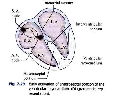
After mid-septal activation from the left to the right direction (Fig. 7.28), the impulse comes down the septum to the apex of the heart and next portions of myocardium that is activated is the anteroseptal region of the ventricular myocardium (Fig. 7.29). The impulse then proceeds along the right and left ventricular walls to the atrioventricular groove.


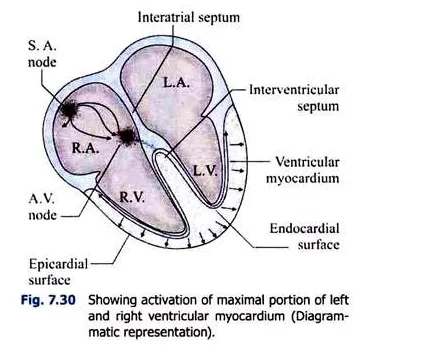
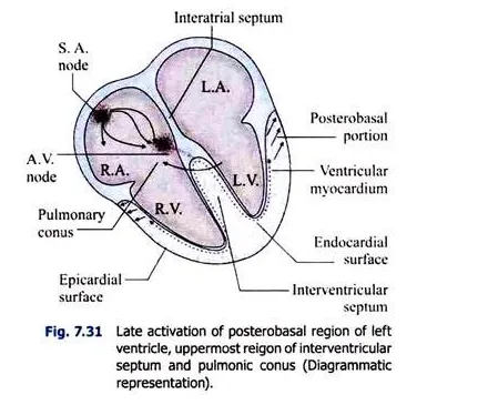
The spread of excitation through the ventricle proceeds from the endocardium to the epicardium and thus the whole of the right and left ventricular walls are depolarised (Fig. 7.30) producing the QRS complex in E.C.G. The portions of the ventricles that are excited lastly are the posterobasal regions of the left ventricle, the pulmonary conus, and the uppermost portion of the interventricular septum (Fig. 7.31).


6. Cell-to-Cell Conduction:
Earlier conception was that there is protoplasmic continuity between cells of the cardiac muscle and thus impulse is transmitted through the intercellular bridges. But electron microscopic studies reveal that there is no such a bridge or protoplasmic continuity, the cells are bounded on all sides by membranes of high resistance.
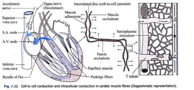
Transmission of impulse through this membrane is impossible. But the intercalated disc which crosses the short axes of the cells offers very low resistance and impulses reaching the intercalated disc are quickly propagated to the cells. So the syncytium-like properties of cardiac muscle are due to presence of low resistance intercalated discs (Fig. 7.32)
7. Intracellular Conduction:
Through the specialised areas of the intercalated discs, the impulse ultimately reaches the cell membrane— sarcolemma. From here, the impulse is transmitted quickly through the transverse tubules (T tubules) —sarcolemmal invagination.
This T tubule passes through the Z-line and here it is in close apposition with the sarcoplasmic reticulum that is present in the area in between the Z-lines. So the impulse from the cell wall is transmitted through the transverse tubules and then through the sarcoplasmic reticulum, and reaches ultimately to the contractile units of the muscle (Fig. 7.32).

If the bundle of His is damaged experimentally in animals or in disease in man, the atria and ventricles are completely dissociated and they beat quite independently. The impulse is transmitted through the right and left branches of the bundle of His to the right and left ventricles respectively. If any branch of the bundle of his is damaged, conduction of the impulses is retarded on the damaged side.
The anterior part of the septal region is excited first, then the apex and lastly the base of the heart. The time taken by the impulse to spread from the bundle of His through its branches to the apex is 0.013 sec. At first the endocardial surface and then the pericardial surface of the ventricles are stimulated. The impulse then spreads from apex to base by the Purkinje fibres.

