Brain
Many regions of the brain can influence the input arriving at lower levels of the nervous system. This descending inhibition can be selective, with different regions of the brain inhibiting certain inputs to the spinal cord. Some regions reduce mechanoreceptive input, and others reduce noxious and warmth inputs. Descending inhibition can also reduce input from the skin while increasing input related to movement.
Prominent regions of influence are those that themselves receive noxious input. For instance, the lateral reticular nuclei of the medulla oblongata cause a constant inhibition of input brought to the spinal cord by the nonmyelinated fibres. In the rat (in which the discovery was first made) descending inhibition can be so effective that a noxious input does not enter the spinal cord. In other words, normally painful stimuli cause no reaction or concern, and there is no change in blood pressure, respiration, or other reflex activities. In these circumstances it seems that pain simply is not felt.
Electrical stimulation of the nucleus ceruleus, a small nucleus with widely ranging axons, and the nucleus raphe magnus, a nucleus in the central reticular formation of the medulla oblongata, inhibits input from noxious stimulation of the skin, and it also inhibits activities of dorsal-horn neurons receiving mechanoreceptive input. Since it was discovered that pain could be obliterated in this manner, attempts have been made to stop the chronic pain of cancer and other conditions by implanting electrodes in relevant parts of the brain so that constant stimulation can inhibit the input coming from the region of the pain.
This system for obliterating or reducing pain can normally be activated by stress. Furthermore, it has always been known that one pain can mask another. A barrage of nerve impulses reaching the brain via the spinothalamic and spinoreticular tracts and causing a moderate degree of pain is stopped when the cells of origin of this tract are inhibited. Experiments have shown that this suppression can be brought about by a more severe pain or by a pain in a larger area of the body, which causes descending inhibition of neurons of the spinal tract. This descending inhibition is the main mechanism of acupuncture.
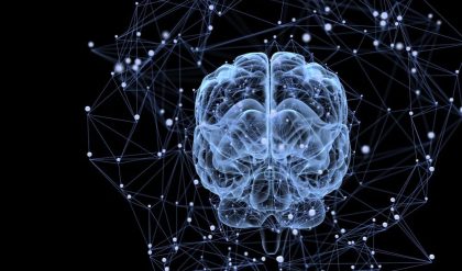
The reticular formation consists of a vast number of small interconnected neurons occupying the central area of the brainstem. Parts of the reticular formation, hypothalamus, and thalamus excite the cerebral hemispheres and keep the cerebral cortex active and alert—partly in response to noxious input. In fact, it may be said that pain reaches consciousness in the thalamus. The thalamus receives noxious input from the spinal cord in two regions, a lateral part called the ventrobasal complex and a medial part consisting of several nuclei. The ventrobasal complex is involved with the accurate temporal and spatial localization of conscious sensation, while the medial nuclei are concerned with the emotional, affective, and autonomic components of pain and other sensations. The ventrobasal nuclei relay impulses to the sensory areas of the postcentral gyrus. Noxious stimuli also cause responses in many areas of the cerebral cortex and the deeper islands of gray matter. This is to be expected, for pain is the least-pure sensation; it startles, it excites, and it has unpleasant qualities. All these aspects of pain are added by different parts of the brain.
Central pain
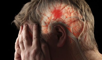
Pain arising within the central nervous system when there is no damage to the body is known as central pain. The most common central pain is due to lesions in or near the thalamus and is called the thalamic syndrome. This condition is characterized by diminution in sensibility as well as severe pain when any stimulus exceeds a certain threshold.
With central pain, there is both spontaneous pain and excessive pain on stimulation of all kinds. Pain may occur in an entire half of the body, affecting even visual and auditory inputs, or it may occur in a restricted region, such as an upper limb and side of the face or a lower limb.
Referred pain
The term referred pain is used to describe pain felt in a region where it does not originate but to which it is referred. It is usually used to describe pain arising in hollow viscera and felt in somatic tissues, such as the body wall. Referred pain is always referred in one direction—from deep to superficial tissues. It is pain referred from an unknown or unfamiliar part of the body to a known or familiar part.
Certain regions of the dorsal horns of the spinal cord receive both nerve fibres from the viscera and nerve fibres reporting noxious events in the skin and musculature. For example, afferent nerve fibres from the heart travel to the same regions as those from the muscles and skin of the chest wall and upper limbs. (Angina pectoris, a spasm of pain in the chest that results from diseased heart tissue, is often referred to the chest and arm.) There is an intermingling of inputs in visceral and somatic tissues, and there is similar convergence in the thalamus. This anatomical arrangement is likely to form the basis of referred pain, although the mere convergence of impulses from viscera and soma onto the same neurons does not alone account for the false localization of visceral pain. It is probable that in sensory areas of the brain, the skin is served by a large number of neurons, the muscles with fewer, and the viscera with least, the visceral representation in the cortex being very small compared with that of the somatic tissues. It is supposed that, as the input to these sensory regions of the cortex usually comes from the skin and body wall, localization of the visceral input will be to these tissues and not to the viscera, the cortical region of which is small and relatively unused.
Changes in the cerebral cortex
Normally, electrical stimulation of the sensory region of the postcentral gyrus does not cause pain. But in many patients who have a painful state on the opposite side of the body, such as an amputation stump or damage to the median nerve of the hand, stimulation of this region reproduces the pain. Pain also arises from stimulation of the white matter deep in the cerebral cortex.
In these cases the character of the sensory region of the cortex changes so that neurons that normally never cause pain when stimulated now invariably produce the pain from which the individual is suffering. Also, the cortical area subserving the limb enlarges. For instance, the sensory area receiving impulses from the opposite lower limb is normally at the upper end of the postcentral gyrus. If there is a painful amputation of the limb, then the area of the cortex in which electrical stimulation induces the pain spreads downward from the normal area to include the trunk area and sometimes the upper limb area; this phenomenon is called phantom pain.


