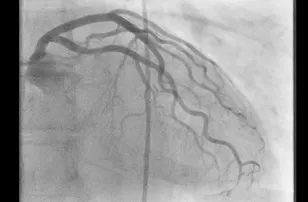
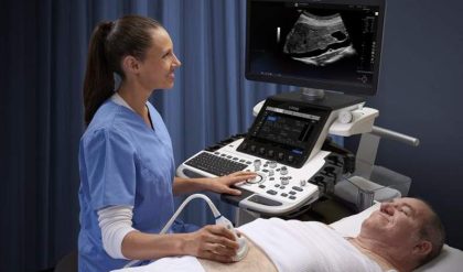
Angiography is an X-ray technique used in the examination of the arteries, veins and organs to diagnose and treat blockages and other blood vessel problems. An interventional radiologist performs the procedure, known as an angiogram. During the angiogram, the radiologist inserts a thin tube, called a catheter, into an artery or vein from an access point. This can be in the groin or the arm; the precise location depends on the area of the body being examined. A substance called a contrast agent is injected to make the blood vessels visible on the X-ray image, e.g. vessels around the knee (Figure 1) 1, vessels around the heart (Figure 2) 2. The agent is later eliminated from the body through the kidneys and urine.

Fig. 1: Angiogram of a blood vessel in the region of the knee.
One of the most common uses of angiograms is to identify blockages in, or narrowing of, blood vessels that could interfere with the normal blood flow in many parts of the body, including the brain, heart, abdomen, and legs. In many cases, the interventional radiologist can treat a blocked blood vessel while the angiogram is performed without the need for surgery.

Fig. 2: Cardiac imaging acquisition using a special low dose program.
Your doctor may recommend an angiogram to diagnose a variety of vascular conditions, including:
· Blockages of the arteries outside the heart (peripheral arterydisease, PAD)
· Enlargement of the arteries (aneurysms)
· Problems with the veins (deep vein thrombosis, DVT) or blood clots in the lungs (pulmonary emboli)
· Problems in the arteries that branch off the aorta (aortic arch conditions)
· Malformed arteries (vascular malformations)
· Kidney artery conditions (renovascular conditions)
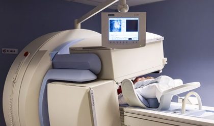
· Angiography is performed with various technologies, for example X-ray using fixed C-arm systems (Figure 3), computed tomography (CT) and magnetic resonance imaging (MRI).

Fig. 3: Fixed C-arm system.
Doctors and manufacturers do all they can to minimize radiation dose, and further reduce the risks involved. Nevertheless, women should always inform their physician or X-ray technologist if there is any possibility that they are pregnant.


