In the second week of prenatal life, the rapidly growing blastocyst (the bundle of cells into which a fertilized ovum divides) flattens into what is called the embryonic disk. The embryonic disk soon acquires three layers: the ectoderm (outer layer), mesoderm (middle layer), and endoderm (inner layer). Within the mesoderm grows the notochord, an axial rod that serves as a temporary backbone. Both the mesoderm and notochord release a chemical that instructs and induces adjacent undifferentiated ectoderm cells to thicken along what will become the dorsal midline of the body, forming the neural plate. The neural plate is composed of neural precursor cells, known as neuroepithelial cells, which develop into the neural tube (see below Morphological development). Neuroepithelial cells then commence to divide, diversify, and give rise to immature neurons and neuroglia, which in turn migrate from the neural tube to their final location. Each neuron forms dendrites and an axon; axons elongate and form branches, the terminals of which form synaptic connections with a select set of target neurons or muscle fibres.
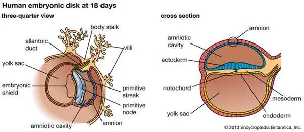
human embryonic developmentDevelopment of the human embryo at 18 days, at disk or shield stage, shown in (left) three-quarter view and (right) cross section.
The remarkable events of this early development involve an orderly migration of billions of neurons, the growth of their axons (many of which extend widely throughout the brain), and the formation of thousands of synapses between individual axons and their target neurons. The migration and growth of neurons are dependent, at least in part, on chemical and physical influences. The growing tips of axons (called growth cones) apparently recognize and respond to various molecular signals, which guide axons and nerve branches to their appropriate targets and eliminate those that try to synapse with inappropriate targets. Once a synaptic connection has been established, a target cell releases a trophic factor (e.g., nerve growth factor) that is essential for the survival of the neuron synapsing with it. Physical guidance cues are involved in contact guidance, or the migration of immature neurons along a scaffold of glial fibres.
In some regions of the developing nervous system, synaptic contacts are not initially precise or stable and are followed later by an ordered reorganization, including the elimination of many cells and synapses. The instability of some synaptic connections persists until a so-called critical period is reached, prior to which environmental influences have a significant role in the proper differentiation of neurons and in fine-tuning many synaptic connections. Following the critical period, synaptic connections become stable and are unlikely to be altered by environmental influences. This suggests that certain skills and sensory activities can be influenced during development (including postnatal life), and for some intellectual skills this adaptability presumably persists into adulthood and late life.
