Bone scan is an imaging technique that uses a radioactive compound to identify areas of healing within the bone. Bone scans work by drawing blood from the patient and tagging it with a bone seeking radiopharmaceutical. This radioactive compound emits gamma radiation. The blood is then returned to the patient intravenously. As the body begins its metabolic activity at the site of the injury, the blood tagged by the radioactive compound is absorbed at the bone and the gamma radiation at the site of the injury can be detected with an external gamma camera. A bone scan can be beneficial in determining injury to the bone within the first 24-48 hours of injury or when the displacement is too small to be detected by an x-ray or CT scan.
Indications for Bone Scans:
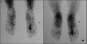
Stress Fracture under Bone Scan
1. Primary and metastatic bone neoplasms.
2. Disease progression or response to therapy.
3. Paget’s disease of bone.
4. Stress and/or occult fractures.
5. Trauma – accidental and non-accidental.
6. Osteomyelitis.
7. Musculoskeletal inflammation or infection.
8. Bone viability (grafts, infarcts, osteonecrosis).
9. Metabolic bone disease.
10. Arthritides.
11. Prosthetic joint loosening and infection.
12. Pain of suspected musculoskeletal etiology.
13. Myositis ossificans.
14. Complex regional pain syndrome (CRPS 1). Reflex sympathetic dystrophy.
15. Abnormal radiographic or laboratory findings.
16. Distribution of osteoblastic activity prior to administration of therapeutic radio-pharmaceuticals for treating bone pain.
Electron Microscopy
The electron microscope is a microscope that can magnify very small details with high resolving power due to the use of electrons as the source of illumination, magnifying at levels up to 2,000,000 times.
Electron microscopy is employed in anatomic pathology to identify organelles within the cells. Its usefulness has been greatly reduced by immunhistochemistry but it is still irreplaceable for the diagnosis of kidney disease, identification of immotile cilia syndrome and many other tasks.
Nuclear Medicine
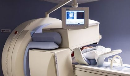
Nuclear medicine on a whole encompasses both the diagnosis and treatment of disease using nuclear properties. In imaging, a radiopharmaceutical is injected to the patient, radiopharmaceuticals are drugs that contain radioactive isotopes, these then decay and emit energy that helps produce the images..
Gamma cameras are used in nuclear medicine to detect regions of biological activity that are often associated with diseases. A short lived isotope, such as 123I is administered to the patient. These isotopes are more readily absorbed by biologically active regions of the body, such as tumors or fracture points in bones.

PET Scan of brain
Positron Emission Tomography
Positron emission tomography (PET) is primarily used to detect diseases of the brain and heart. Similarly to nuclear medicine, a short-lived isotope, such as 18F, is incorporated into a substance used by the body such as glucose which is absorbed by the tumor of interest. PET scans are often viewed alongside computed tomography scans, which can be performed on the same equipment without moving the patient. This allows the tumors detected by the PET scan to be viewed next to the rest of the patient’s anatomy detected by the CT scan.
Single Photon Emission Computed Tomography
Single Photon Emission Computed Tomography (SPECT) is a widely used imaging technique in nuclear medicine for the visualization of organs, such as the bones, heart and brain, as well as for the detection of tumors. Because of its capability to visualize and quantify changes in the cerebral blood flow and neurotransmitter system, it has important use in the differential diagnosis of neurological and psychiatric diseases.
Optoacoustic Imaging
Also known as Photoacoustic Imaging, is an upcoming biomedical imaging modality availing the benefits of optical resolution and acoustic depth of penetration. With its capacity to offer structural, functional, molecular and kinetic information making use of either endogenous contrast agents like hemoglobin, lipid, melanin and water or a variety of exogenous contrast agents or both, Optoacoustic imaging has demonstrated promising potential in a wide range of preclinical and clinical applications.
Clinical applications of optoacoustic imaging include:
· Breast imaging
· Dermatologic Imaging
· Pilosebaceous units
· Skin cancer
· Inflammatory skin diseases
· Vascular Imaging
· Cutaneous miscrovasculature
· Vascular Dysfunction
· Wound Imaging
· Carotid Vessel Imaging
· Musculoskeletal Imaging
· Gastrointestinal Imaging
· Adipose Tissue Imaging

Comments are closed.