Molecular imaging uses small amounts of radioactive markers, called radiopharmaceuticals, in the visualization and diagnosis of disease, including many types of cancer, heart disease, neurological disorders and other abnormalities in the body.
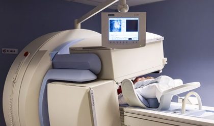
Depending on the type of examination, the radiopharmaceutical is either injected in liquid form into a vein or swallowed by the patient, or it can be inhaled as a gas. The radiopharmaceuticals accumulate in the organ or area of the body being examined, where they give off energy in the form of gamma rays. This energy is detected by positron emission tomography (PET) or single photo emission (SPECT) scanner (Figure 1) 1. A computer is used to calculate the amount of radiopharmaceutical absorbed by the body and special images are produced providing details on both the structure and function of organs and tissues.
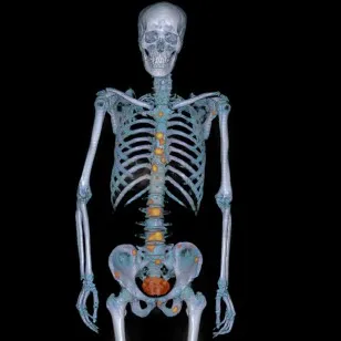
Fig. 1: SPECT image
Molecular imaging procedures allow visualization of the structure and function of organs, tissue, bones or other systems of the body, e.g. heart blood flow and function, and kidney and lung function. Furthermore, molecular imaging scans are performed for a wide range of purposes from locating lymph nodes before surgery in patients with breast cancer or melanoma to evaluating bone fractures, infections and tumors or investigating abnormalities in the brain, such as seizures and memory loss.
Radiopharmaceuticals are also used to treat cancer and metastases, medical conditions affecting the thyroid gland, certain blood disorders and adrenal gland tumors in adults and nerve tissue tumors in children.
The amount of radioactive material used during these examinations depends on the individual procedure. The risk to the patient is comparably low, and experts consider the risks to be far outweighed by the benefits of an accurate diagnosis and treatment. Doctors and manufacturers know about these risks and are working together to minimize radiation dose. Nevertheless, women should always inform their physician or X-ray technologist if there is any possibility that they are pregnant.
Combined Modalities

Fig. 1: PET image
Molecular imaging offers physicians a unique insight into the workings of the human body at a cellular and molecular level, enabling them to diagnose and to characterize potential disease at a very early stage. To correlate the biological processes with anatomical location in the body, molecular imaging devices are integrated with CT and MRI scanners (Figure 1) 1. Computers are used to fuse the biological and anatomical images together to help doctors make better diagnostic and therapeutic decisions.
PET & CT exposes the patient to a small amount of ionizing radiation. The exact amount varies according to the type and length of the procedure and this in turn is associated with a low risk to the patient. Doctors and manufacturers know about these risks and do all they can to minimize radiation dose, keeping the exposure time to a minimum. These risks, however, are remote and experts consider them to be far outweighed by the benefits of an accurate diagnosis and treatment. Nevertheless, women should always inform their physician or X-ray technologist if there is any possibility that they are pregnant.

Fig. 2: PET-MR image
In 2010, a new hybrid imaging modality was introduced: known as MR-PET or PET/MR. It combines the technologies of both MR and PET. Across the globe, there are around 100 hybrid MR-PET systems installed, most of them in university hospitals, research institutes and larger imaging centers. In order to generate a whole-body PET image, a small amount of a radioactive substance is injected into the patient’s bloodstream prior to the scan. At the same time, a high-resolution magnetic resonance imaging (MRI) exam is performed which does not utilize any additional ionizing radiation.
Where both PET and MRI imaging are required to resolve diagnostic questions, the MR-PET has a key benefit: Two examinations are performed in a single scan. This saves time: The patient needs just one examination, and the physician receives both results at the same time. Furthermore, MRI can provide additional soft tissue in contrast to CT.

Comments are closed.