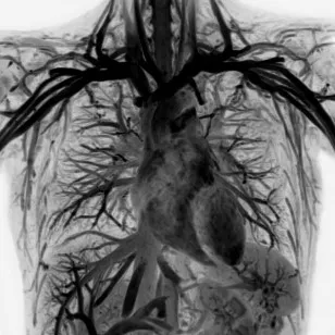
MRI systems use a powerful magnetic field and radiofrequency pulses to produce detailed images of the body’s internal structures as cross-sectional images or slices (Figure 1). Without exposing the patient or staff to ionizing radiation (X-rays), MRI provides high quality images with excellent contrast detail of soft tissue and anatomic structures such as gray and white matter in the brain. It’s used in a wide range of examinations from brain tumors and inflammation of the spine to slipped discs, assessing blood flow and functioning of the heart. MRI does not emit any ionizing radiation.

Fig. 1: MRI provides excellent high contrast definition, as shown in this image of the thorax.
Many diseases, such as certain brain tumors, can be visualized using MRI because of a high contrast definition, which does not always require contrast agents to produce detailed images of blood vessels. MRI scanners can image a wide range of body parts including injuries of the joints, the blood vessels, the breast, as well as abdominal and pelvic organs such as the liver or reproductive organs.
The MRI system consists of a very powerful superconducting magnet that creates a static magnetic field, smaller ‘gradient’ magnets that allow the magnetic field to be very precisely altered and designated coils for specific body parts that emit radiowaves.
During the examination, the gradient coils are used to ‘focus’ the magnetic field on the part of the body to be scanned. The radio signal is turned on and off and the energy absorbed by different atoms is reflected back out of the body. The coil measures these radio waves and the computer then calculates the way they have been absorbed or reflected to compile the cross-sectional images. The tapping sound heard during the examination is caused when gradient magnets are switched on and off.
MRI scanners can acquire direct views of the body in almost any orientation and without exposing the patient or staff to ionizing radiation (X-rays). Care must be taken, however, in patients with implants, because they might be affected by the strong magnetic field. Patients with common pacemakers, for example, can generally not have an MRI scan. There is also a slight risk of allergic reaction by some patients to contrast agents, if used.


