Computed tomography (CT) scanners have been available since the mid-1970s and have revolutionized medical imaging. Today, millions of scans are performed worldwide every year for different clinical questions in a variety of clinical fields. In emergencies for example, CT is widely used as it delivers detailed information fast, which is essential for appropriate treatment decisions.
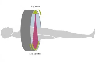
Fig. 1: The principle of computed tomography with an X-ray source and detector unit rotating synchronously around the patient. Data are acquired continuously during rotation.
The most prominent part of a CT scanner is the gantry – a circular, rotating frame with an X-ray tube mounted on one side and a detector on the opposite side. A fan-shaped beam of X-rays is created as the rotating frame spins the X-ray tube and detector around the patient. As the scanner rotates, several thousand sectional views of the patient’s body are generated in one rotation, which result in reconstructed cross-sectional images of the body (Figure 1)1. Using on these data, it is possible to create a 3D visualization and views from different angles.

Fig. 2: Modern CT scans provide very detailed images, for example of blood vessels and internal organs, by using relatively low radiation doses. This CT scan of the whole thorax and abdomen was performed in less than 1 sec with only 20 mL of contrast media, and a radiation dose of 2.32 mSv.
CT scans provide far more detailed images than conventional X-ray imaging, especially in the case of blood vessels and soft tissue such as internal organs and muscles. A part of the energy of the X-ray beam is absorbed when it passes through the body. This process is described as the attenuation of the X-ray beam. Just as with an X-ray film, this attenuation depends on the tissue. Bone appears white since the attenuation of bone is very high. For air the opposite is true, so air appears black. With modern CT scans, you can even apply colorcoding to the dataset as seen in Figure 2. Scans can be completed in seconds or even fractions of a second. For some CT scans, a special contrast agent is injected into a vein before the scan as this allows further assessment of the organs and vessels (Figure 2)2. These preparations for the examination may require additional time. A CT scanner can image any part of the body including the heart, lungs and abdomen. CT scans are also invaluable in assessing skeletal injuries, as even very tiny bones are shown clearly.
During the scan, the patient lies on a table that is moved through the ring-shaped gantry of the CT scanner for the examination. CT scanning is not painful and is safe for people with pacemakers.
CT scans are valuable in emergencies because they are able to provide information very quickly. This is important, for example when assessing strokes, brain injuries, heart disease, and internal injuries. In addition, the short duration of the scanning process benefits patients who are not easily able to keep still, such as children. CT imaging is a very important tool to diagnose cancer and to obtain additional information for different clinical questions. A CT scan usually requires a higher radiation exposure dose than a conventional radiography examination. However, a CT scan delivers more detailed information. Doctors and manufacturers do all they can to minimize radiation dose. As with conventional X-ray imaging, CT scans are not recommended for pregnant women unless the examination is absolutely necessary.

