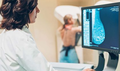
When imaging with X-rays, an X-ray beam produced by a so-called X-ray tube passes through the body. On it’s way through the body, parts of the energy of the X-ray beam are absorbed. This process is described as attenuation of the X-ray beam. On the opposite side of the body, detectors or a film capture the attenuated X-rays, resulting in a clinical image. In conventional radiography, one 2D image is produced. In Computed Tomography, the tube and the detector are both rotating around the body during the examination so that multiple images can be acquired, resulting in a 3D visualization.
The most common methods of X-ray in medical imaging are X-ray radiography, computed tomography (CT), mammography, angiography and fluoroscopy.

Fig. 1: Chart comparing typical effective doses of medical examinations with the average natural background radiation.
Different organs and tissues have a different sensitivity to radiation. This is why the actual risk to the body from X-ray procedures varies depending on the part of the body being X-rayed. “Effective dose” is a parameter of the dose absorbed by the entire body that takes account of these differing sensitivities.
Doctors and manufacturers are well aware of the risks and do everything possible to minimize radiation dose. Guided by technical standards that are set and continually updated by national and international radiology protection councils, they take special care during X-ray examinations to use the lowest radiation dose possible while producing the images for. Advanced X-ray systems contain special features that help reduce the radiation dose. For example there are technologies developed to ensure that those parts of a patient’s body not being imaged receive no or only minimal radiation exposure.


